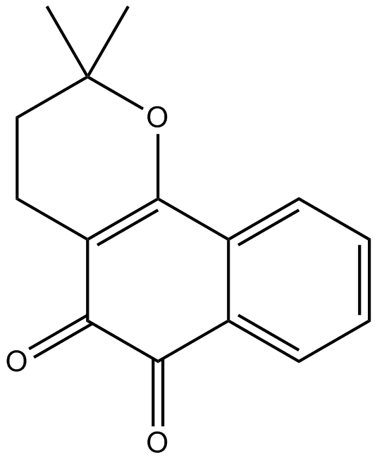
β-Lapachone

(Synonyms: 3,4-二氢-2,2-二甲基-2H-萘并[1,2-B]吡喃-5,6-二酮,ARQ-501; NSC-26326) 目录号 : GC10734
β-Lapachone是一种邻萘醌天然产物,具有抗炎、抗菌及抗锥虫等多种生物活性,且能抑制拓扑异构酶I并诱导NAD(P)H:醌氧化还原酶1。

Cas No.:4707-32-8
Sample solution is provided at 25 µL, 10mM.
β-Lapachone is an ortho-naphthoquinone natural product, promoting several biological effects, such as anti-inflammatory, antibacterial, and anti-Trypanosoma, and β-Lapachone can inhibit topoisomerase I and induce NAD(P)H: quinone oxidoreductase 1[1-3].
In vitro, after colon cancer cells SW480 cells were transiently exposed to β-Lapachone (2.5 or 5µM) for 4h, β-Lapachone induced primarily S-phase arrest in SW480 cells in a concentration-dependent manner[4]. Treatment of human hepatocellular carcinoma SK-Hep1 cells with different concentrations of β-Lapachone (1, 2, 4μM) for 18h showed that β-Lapachone increased the morphological appearance of dying cells in a dose-dependent manner, and cell viability was decreased in β-Lapachone-treated cells; furthermore, β-Lapachone also increased propidium iodide (PI) uptake (loss of plasma membrane integrity) in a dose-dependent manner[5].
In vivo, male Balb/c mice fed a diet containing β-Lapachone (0.066%) for 2 weeks before cisplatin injection (18mg/kg; i.p.), β-Lapachone markedly decreased cisplatin-induced renal cell apoptosis and DNA double-strand breaks (DSBs) formation, which was accompanied by enhanced basal expression of SIRTuin1, and the β-Lapachone + cisplatin group showed the highest Mre11-Rad50-Nbs1 (MRN) complex expression[6]. Log-phase A549 cells were injected subcutaneously into the flanks of athymic nude mice, followed by β-Lapachone (30mg/kg) injection through the tail vein, and β-Lapachone demonstrated potent in vivo antitumor activity with Tumor growth inhibition (TGI) = 70.1% and tumor proliferation rate (T/C) = 29.5%[7].
References:
[1] Gong Q, Hu J, Wang P, et al. A comprehensive review on β-lapachone: Mechanisms, structural modifications, and therapeutic potentials. Eur J Med Chem. 2021;210:112962.
[2] Ferraz da Costa DC, Pereira Rangel L, Martins-Dinis MMDDC, et al. Anticancer Potential of Resveratrol, β-Lapachone and Their Analogues. Molecules. 2020;25(4):893.
[3] Gomes CL, de Albuquerque Wanderley Sales V, Gomes de Melo C, et al. Beta-lapachone: Natural occurrence, physicochemical properties, biological activities, toxicity and synthesis. Phytochemistry. 2021;186:112713.
[4] Huang L, Pardee AB. beta-lapachone induces cell cycle arrest and apoptosis in human colon cancer cells. Mol Med. 1999;5(11):711-720.
[5] Park EJ, Min KJ, Lee TJ, Yoo YH, Kim YS, Kwon TK. β-Lapachone induces programmed necrosis through the RIP1-PARP-AIF-dependent pathway in human hepatocellular carcinoma SK-Hep1 cells. Cell Death Dis. 2014;5(5):e1230.
[6] Kim TW, Kim YJ, Kim HT, et al. β-Lapachone enhances Mre11-Rad50-Nbs1 complex expression in cisplatin-induced nephrotoxicity. Pharmacol Rep. 2016;68(1):27-31.
[7] Li X, Bian J, Wang N, et al. Novel naphtho[2,1-d]oxazole-4,5-diones as NQO1 substrates with improved aqueous solubility: Design, synthesis, and in vivo antitumor evaluation. Bioorg Med Chem. 2016;24(5):1006-1013.
β-Lapachone是一种邻萘醌天然产物,具有抗炎、抗菌及抗锥虫等多种生物活性,且能抑制拓扑异构酶I并诱导NAD(P)H:醌氧化还原酶1[1-3]。
在体外,将结肠癌细胞SW480短暂暴露于β-Lapachone(2.5或5µM)4小时后,β-Lapachone以浓度依赖性方式主要诱导SW480细胞发生S期阻滞[4]。采用不同浓度β-Lapachone (1、2、4μM)处理人肝细胞癌SK-Hep1细胞18小时的结果显示,β-Lapachone以剂量依赖性方式增加死亡细胞的形态学变化,β-Lapachone处理组细胞活力下降;此外,β-Lapachone亦以剂量依赖性方式增加碘化丙啶(PI)摄取(质膜完整性丧失)[5]。
在体内,雄性Balb/c小鼠于顺铂注射(18mg/kg;腹腔注射)前2周开始饲喂含β-Lapachone (0.066%)的饲料,β-Lapachone显著减少顺铂诱导的肾细胞凋亡和DNA双链断裂(DSBs)形成,伴随SIRTuin1基础表达上调,且β-Lapachone加顺铂组Mre11-Rad50-Nbs1(MRN)复合物表达最高[6]。将处于对数生长期的A549细胞皮下接种于无胸腺裸鼠侧腹,随后经尾静脉注射β-Lapachone (30mg/kg),β-Lapachone表现出强效的体内抗肿瘤活性,肿瘤生长抑制率(TGI)=70.1%,肿瘤增殖率(T/C)=29.5%[7]。
| Cell experiment [1]: | |
Cell lines | SW480 cells, SW620 cells, DLD1 cells |
Preparation Method | Cells were seeded into 60mm dishes at a density of 7×10⁵ per dish. Colon cancer cells, SW480, SW620, and DLD1 cells, were transiently exposed to β-Lapachone (2.5 or 5µM) for 4h, further incubated in drug-free medium for 20h, and then analyzed for cell cycle distribution by flow-cytometric analysis. Cells were then harvested by trypsinization, and incubated for 30min at room temperature in staining solution consisting of propidium iodide (50µg/ml), sodium citrate (0.1%), Triton X-100 (0.1%), and DNase-free RNase (20µg/ml). Stained cells were then analyzed for DNA content by flow cytometry. |
Reaction Conditions | 2.5 and 5µM; 4h |
Applications | β-Lapachone induced primarily S-phase arrest in SW480 cells, late S- and G2/M-phase arrest in SW620 cells, and early S-phase arrest in DLD1 cells. The cell cycle alterations were concentration-dependent. At 2.5µM, β-Lapachone induced S-phase arrest in all cell lines. When the concentration was increased to 5µM, more SW480 cells were arrested in S phase (53.1% at 5µM as compared to 43% at 2.5µM and 34.5% at control), more SW620 cells were arrested in S/G2 phase (93.7% at 5µM as compared to 64.5% at 2.5µM and 56.6% at control), and more DLD1 cells were arrested in early S phase. |
| Animal experiment [2]: | |
Animal models | Male Balb/c mice |
Preparation Method | After 1 week of acclimation, the mice were randomly allocated to one of the following groups (5 per group): control, β-Lapachone, cisplatin (18mg/kg, i.p.), and β-Lapachone + cisplatin (18mg/kg, i.p.). The β-Lapachone groups were fed a diet containing the drug (0.066%) for 2 weeks before cisplatin injection. All mice were sacrificed under carbon dioxide anesthesia 3 days after cisplatin injection. The blood samples obtained from the retro-orbital plexus were subjected to serum blood urea nitrogen (BUN) and creatinine (CRE) analyses. Half of the kidney was quickly removed for histopathological and immunohistochemical (IHC) studies. The other half was stored at -70°C until the western blot assay. |
Dosage form | 0.066%; orally |
Applications | In the cisplatin-alone group, renal function was disrupted, and Mre11-Rad50-Nbs1 (MRN) complex expression increased. β-Lapachone co-treatment attenuated cisplatin-induced pathologic alterations. Although β-Lapachone markedly decreased cisplatin-induced renal cell apoptosis and DNA double-strand breaks (DSBs) formation, the β-Lapachone + cisplatin group showed the highest MRN complex expression. Moreover, β-Lapachone treatment increased the basal expression level of the MRN complex, which was accompanied by enhanced basal expression of SIRTuin1. |
References: | |
| Cas No. | 4707-32-8 | SDF | |
| 别名 | 3,4-二氢-2,2-二甲基-2H-萘并[1,2-B]吡喃-5,6-二酮,ARQ-501; NSC-26326 | ||
| 化学名 | 2,2-dimethyl-3,4-dihydrobenzo[h]chromene-5,6-dione | ||
| Canonical SMILES | CC1(CCC2=C(O1)C3=CC=CC=C3C(=O)C2=O)C | ||
| 分子式 | C15H14O3 | 分子量 | 242.27 |
| 溶解度 | ≥ 10.85mg/mL in DMSO | 储存条件 | Store at -20° C |
| General tips | 请根据产品在不同溶剂中的溶解度选择合适的溶剂配制储备液;一旦配成溶液,请分装保存,避免反复冻融造成的产品失效。 储备液的保存方式和期限:-80°C 储存时,请在 6 个月内使用,-20°C 储存时,请在 1 个月内使用。 为了提高溶解度,请将管子加热至37℃,然后在超声波浴中震荡一段时间。 |
||
| Shipping Condition | 评估样品解决方案:配备蓝冰进行发货。所有其他可用尺寸:配备RT,或根据请求配备蓝冰。 | ||
| 制备储备液 | |||
 |
1 mg | 5 mg | 10 mg |
| 1 mM | 4.1276 mL | 20.6381 mL | 41.2763 mL |
| 5 mM | 0.8255 mL | 4.1276 mL | 8.2553 mL |
| 10 mM | 0.4128 mL | 2.0638 mL | 4.1276 mL |
| 第一步:请输入基本实验信息(考虑到实验过程中的损耗,建议多配一只动物的药量) | ||||||||||
| 给药剂量 | mg/kg |  |
动物平均体重 | g |  |
每只动物给药体积 | ul |  |
动物数量 | 只 |
| 第二步:请输入动物体内配方组成(配方适用于不溶于水的药物;不同批次药物配方比例不同,请联系GLPBIO为您提供正确的澄清溶液配方) | ||||||||||
| % DMSO % % Tween 80 % saline | ||||||||||
| 计算重置 | ||||||||||
计算结果:
工作液浓度: mg/ml;
DMSO母液配制方法: mg 药物溶于 μL DMSO溶液(母液浓度 mg/mL,
体内配方配制方法:取 μL DMSO母液,加入 μL PEG300,混匀澄清后加入μL Tween 80,混匀澄清后加入 μL saline,混匀澄清。
1. 首先保证母液是澄清的;
2.
一定要按照顺序依次将溶剂加入,进行下一步操作之前必须保证上一步操作得到的是澄清的溶液,可采用涡旋、超声或水浴加热等物理方法助溶。
3. 以上所有助溶剂都可在 GlpBio 网站选购。
Quality Control & SDS
- View current batch:
- Purity: >98.00%
- COA (Certificate Of Analysis)
- SDS (Safety Data Sheet)
- Datasheet


