CFDA-SE

(Synonyms: 5(6)-羧基二乙酸荧光素琥珀酰亚胺酯,5(6)-Carboxyfluorescein diacetate succinimidyl ester; 5(6)-CFDA N-succinmidyl ester) 目录号 : GC14056
CFDA-SE(carboxycein diacetate, Succinimidyl Ester)具有细胞膜渗透性和荧光性,在可视化追踪或细胞增殖检测中得到广泛应用。

Cas No.:150347-59-4
Sample solution is provided at 25 µL, 10mM.
CFDA-SE, full name carboxycein diacetate, Succinimidyl Ester, with cell membrane permeability, is a fluorescent dye CFDA-SE widely used for viable cell tracing or cell proliferation detection[1].The fluorescence of CFDA-SE -labeled cells was very uniform and stable, and the fluorescence could be evenly distributed to the two progeny cells [2]. CFDA-SE can deliver strong green fluorescence,Ex=494 nm,Em=521nm.
In Human erythroleukaemic cell line K562, and other cell lines, although all cells are labeled with relatively high fluorescence intensity by CFDA-SE, the efficiency of labeling is highly variable[3].Electron dense vesicles are seen at the ultrastructural level in labeled cells. Discrete vesicular labeling can also be observed in whole cell mounts viewed with fluorescence microscopy. Whole cells retain good label for 6 weeks. CFDA-SE labeling is relatively easy, nontoxic to cells and nonradiocactive[4].
In vitro lymphocyte cell proliferation analysis by CFDA-SE method is a reliable and practical choice for the assessment of mitogenic T lymphocyte responses in yet unclassified PID patients for targeting further genetical analyses[5].When NM is detected by flow cytometry, FL1 detection channel CFDA-SE -labeled cells can be adopted, and fluorescence microscopy can also be used to observe CFDA-SE -labeled cells without staining adjacent cells [6].
CFDA-SE has been reported to be effective in labeling CD34+ cells, CD34+ cells isolated from fetal liver or thymus were labeled with CFDA-SE and were injected into a human thymus grafted subcutaneously in the RAG-2 / IL-2Rγ / mice. One to 4 weeks later the CFDA-SE label was found not only in T cells but also in CD123+/high CD4+CD45RA+ pDC2, indicating that the CD34+ cells can develop into pDC2 within a thymus. In addition to pDC2, CFDA-SE -labeled dendritic cells with a mature phenotype, determined by the cell surface markers CD11c, CD83, and CD80, were found in the injected human thymus graft. pDC2 was not found in the periphery of mice carrying a human thymic graft, indicating that the intrathymic pDC2 failed to emigrate from the thymus[7]. In C57BL/6 mice,CFDA-SE, at 80 times the concentration used for in vitro labeling, was nontoxic and labeled randomly approximately 15% of thymocytes 24 h after injection. The turnover rate of labeled thymic emigrants in the lymph nodes was in the order of 21 days. Thus, CFDA-SE may serve as a powerful tool in relatively long-term migration studies[8].
References:
[1]: Chen JC, Chang ML, et,al. A kinetic study of the murine mixed lymphocyte reaction by 5,6-carboxyfluorescein diacetate succinimidyl ester labeling. J Immunol Methods. 2003 Aug;279(1-2):123-33. doi: 10.1016/s0022-1759(03)00236-9. PMID: 12969553.
[2]: Lyons AB, Blake SJ, et,al. Flow cytometric analysis of cell division by dilution of CFSE and related dyes. Curr Protoc Cytom. 2013;Chapter 9:Unit9.11. doi: 10.1002/0471142956.cy0911s64. PMID: 23546777.
[3]: Wang XQ, Duan XM, et,al. Carboxyfluorescein diacetate succinimidyl ester fluorescent dye for cell labeling. Acta Biochim Biophys Sin (Shanghai). 2005 Jun;37(6):379-85. doi: 10.1111/j.1745-7270.2005.00051.x. PMID: 15944752.
[4]: Gruber HE, Leslie KP, et,al. Optimization of 5-(and-6)-carboxyfluorescein diacetate succinimidyl ester for labeling human intervertebral disc cells in vitro. Biotech Histochem. 2000 May;75(3):118-23. doi: 10.3109/10520290009066489. PMID: 10950173.
[5]: Azarsiz E, Karaca N, et,al. In vitro T lymphocyte proliferation by carboxyfluorescein diacetate succinimidyl ester method is helpful in diagnosing and managing primary immunodeficiencies. J Clin Lab Anal. 2018 Jan;32(1):e22216. doi: 10.1002/jcla.22216. Epub 2017 Apr 6. PMID: 28383134; PMCID: PMC6816938.
[6]: Li X, Dancausse H, et,al. Labeling Schwann cells with CFSE-an in vitro and in vivo study. J Neurosci Methods. 2003 May 30;125(1-2):83-91. doi: 10.1016/s0165-0270(03)00044-x. PMID: 12763234.
[7]: Weijer K, Uittenbogaart CH, et,al. Intrathymic and extrathymic development of human plasmacytoid dendritic cell precursors in vivo. Blood. 2002 Apr 15;99(8):2752-9. doi: 10.1182/blood.v99.8.2752. PMID: 11929763.
[8]: Graziano M, St-Pierre Y, et,al. The fate of thymocytes labeled in vivo with CFSE. Exp Cell Res. 1998 Apr 10;240(1):75-85. doi: 10.1006/excr.1997.3900. PMID: 9570923.
CFDA-SE(carboxycein diacetate, Succinimidyl Ester)具有细胞膜渗透性和荧光性,在可视化追踪或细胞增殖检测中得到广泛应用[1]。CFDA-SE 标记的细胞的荧光非常均一和稳定,并且荧光可以均匀地分布到两个后代细胞中[2]。CFDA-SE 可以释放强烈的绿色荧光,Ex=494 nm,Em=521nm。
在人类红细胞白血病细胞系K562和其他细胞系中,虽然所有细胞都被 CFDA-SE 标记了相对较高的荧光强度,但标记效率高度可变[3]。在标记的细胞的超微结构水平上可以看到电子稠密的小泡。在荧光显微镜下观察的整个细胞中也可以观察到离散的小泡标记。整个细胞可以保持好的标记效果长达6周。CFDA-SE 标记相对容易,对细胞无毒,并且无放射性[4]。
采用 CFDA-SE 法进行体外淋巴细胞增殖分析是评估未归类PID患者有丝分裂T淋巴细胞反应以进行进一步遗传分析的可靠和实用选择[5]。在流式细胞术中检测 NM 时,可以采用 FL1 检测通道的 CFDA-SE 标记细胞,并且荧光显微镜也可以用于在未染色相邻细胞中观察 CFDA-SE 标记细胞[6]。
CFDA-SE 对标记 CD34+细胞具有效果,从胎儿肝脏或胸腺中分离的 CD34 + 细胞以 CFDA-SE 标记,并被注射到人类胸腺的移植皮下的 RAG-2 / IL-2Rγ / 小鼠中。1-4周后,CFDA-SE 标记不仅在 T 细胞中发现,而且在 CD123+/high CD4+CD45RA+ pDC2 中也发现,表明 CD34+细胞可以在胸腺中发展成为 pDC2。除了 pDC2,注射入人类胸腺移植体中还发现表面标志物为 CD11c、CD83和CD80 的成熟表型的 CFDA-SE 标记树突状细胞。在携带人类胸腺移植体的小鼠的外围没有发现 pDC2,表明胸腺内pDC2未能从胸腺移出[7]。在 C57BL/6小鼠中,CFDA-SE 在体外标记时的浓度的80倍,不具有毒性,并且24小时后随机标记了大约15%的胸腺细胞。标记的胸腺移民在淋巴结中的周转率为21天。因此,CFDA-SE 可以作为相对长期的迁移研究的有力工具[8]。
|
Protocol for Cell labeling and counting with CFDA-SE [1]: |
|
|
Cells were counted and stained with CFDA-SE, as follows:
This protocol only provides a guideline, and should be modified according to your specific needs. |
|
|
References: [1]. Urbani S, Caporale R,et,al. Use of CFDA-SE for evaluating the in vitro proliferation pattern of human mesenchymal stem cells. Cytotherapy. 2006;8(3):243-53. doi: 10.1080/14653240600735834. PMID: 16793733. |
|
|
Cell experiment [1]: |
|
|
Cell lines |
Human erythroleukaemic cell line K562, mouse lymphoma cell line YAC-1, human mammary cancer cell line MCF-7 and human melanoma cell line A375 |
|
Preparation method |
The 5 mM CFDA-SE stock in DMSO was diluted to different concentrations (2 μM, 3 μM, 4 μM, 5 μM, 10 μM and 20 μM) in PBS with a total volume of 1 ml. Cells were added to equal volume of CFDA-SE with different concentrations and incubated at 37 C for 5, 6, 7, 8, 10 and 15 min with agitation. |
|
Reaction Conditions |
1, 1.5, 2, 2.5, 5 and 10 µM CFDA-SE |
|
Applications |
CFDA-SE at 2.5 μM stained more than 95% of the cells on all cell lines tested, and dose-dependently increased fluorescence intensity of stained cells. The optimal concentration for K562 and YAC-1 was found to be 2.5 μM, while the optimal concentrations for A375 and MCF-7 were found to be 5 μM and 10 μM, respectively. Within the 6 h experiment period, no cytotoxicity related to CFDA-SE was observed. |
|
Animal experiment [2]: |
|
|
Animal models |
C57BL/6 mice, aged 5 ~ 8 weeks |
|
Preparation method |
Injected CFDA-SE into thymic lobe |
|
Dosage form |
The concentration of CFDA-SE is 10 μM |
|
Applications |
CFDA-SE, at 80 times the concentration used for in vitro labeling, was nontoxic and labeled randomly approximately 15% of thymocytes 24 h after injection. The turnover rate of labeled thymic emigrants in the lymph nodes was in the order of 21 days. Thus, CFDA-SE may serve as a powerful tool in relatively long-term migration studies. |
|
References: [1]: Wang XQ, Duan XM,et,al. Carboxyfluorescein diacetate succinimidyl ester fluorescent dye for cell labeling. Acta Biochim Biophys Sin (Shanghai). 2005 Jun;37(6):379-85. doi: 10.1111/j.1745-7270.2005.00051.x. PMID: 15944752. [2]: Graziano M, St-Pierre Y, et,al. The fate of thymocytes labeled in vivo with CFSE. Exp Cell Res. 1998 Apr 10;240(1):75-85. doi: 10.1006/excr.1997.3900. PMID: 9570923. |
|
| Cas No. | 150347-59-4 | SDF | |
| 别名 | 5(6)-羧基二乙酸荧光素琥珀酰亚胺酯,5(6)-Carboxyfluorescein diacetate succinimidyl ester; 5(6)-CFDA N-succinmidyl ester | ||
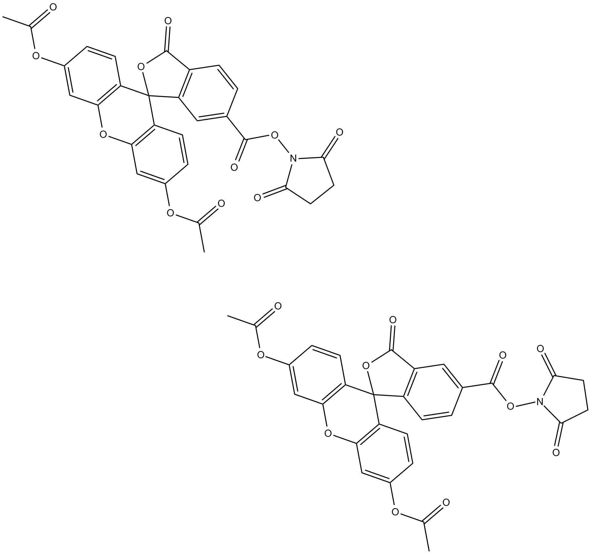
| 化学名 | 5-(((2,5-dioxopyrrolidin-1-yl)oxy)carbonyl)-3-oxo-3H-spiro[isobenzofuran-1,9'-xanthene]-3',6'-diyl diacetate | ||
| Canonical SMILES | O=C1N(OC(C2=CC=C(C3(C(C=CC(OC(C)=O)=C4)=C4OC5=C3C=CC(OC(C)=O)=C5)OC6=O)C6=C2)=O)C(CC1)=O | ||
| 分子式 | C29H19NO11 | 分子量 | 557.46 |
| 溶解度 | ≥ 37.2mg/mL in DMSO with ultrasonic | 储存条件 | Store at -20°C, protect from light |
| General tips | 请根据产品在不同溶剂中的溶解度选择合适的溶剂配制储备液;一旦配成溶液,请分装保存,避免反复冻融造成的产品失效。 储备液的保存方式和期限:-80°C 储存时,请在 6 个月内使用,-20°C 储存时,请在 1 个月内使用。 为了提高溶解度,请将管子加热至37℃,然后在超声波浴中震荡一段时间。 |
||
| Shipping Condition | 评估样品解决方案:配备蓝冰进行发货。所有其他可用尺寸:配备RT,或根据请求配备蓝冰。 | ||
| 制备储备液 | |||
 |
1 mg | 5 mg | 10 mg |
| 1 mM | 1.7939 mL | 8.9693 mL | 17.9385 mL |
| 5 mM | 0.3588 mL | 1.7939 mL | 3.5877 mL |
| 10 mM | 0.1794 mL | 0.8969 mL | 1.7939 mL |
| 第一步:请输入基本实验信息(考虑到实验过程中的损耗,建议多配一只动物的药量) | ||||||||||
| 给药剂量 | mg/kg |  |
动物平均体重 | g |  |
每只动物给药体积 | ul |  |
动物数量 | 只 |
| 第二步:请输入动物体内配方组成(配方适用于不溶于水的药物;不同批次药物配方比例不同,请联系GLPBIO为您提供正确的澄清溶液配方) | ||||||||||
| % DMSO % % Tween 80 % saline | ||||||||||
| 计算重置 | ||||||||||
计算结果:
工作液浓度: mg/ml;
DMSO母液配制方法: mg 药物溶于 μL DMSO溶液(母液浓度 mg/mL,
体内配方配制方法:取 μL DMSO母液,加入 μL PEG300,混匀澄清后加入μL Tween 80,混匀澄清后加入 μL saline,混匀澄清。
1. 首先保证母液是澄清的;
2.
一定要按照顺序依次将溶剂加入,进行下一步操作之前必须保证上一步操作得到的是澄清的溶液,可采用涡旋、超声或水浴加热等物理方法助溶。
3. 以上所有助溶剂都可在 GlpBio 网站选购。
Quality Control & SDS
- View current batch:
- Purity: >98.00%
- COA (Certificate Of Analysis)
- SDS (Safety Data Sheet)
- Datasheet


