
IF 16
PMID: 39846120 ↗Hoechst 33342
目录号: GC63515纯度: >98.00%同义词: 赫斯特荧光染料33342,bisBenzimide H 33342; HOE 33342
核酸染色剂Hoechst 33342 (Ex/Em: 350/461 nm) 常被用作细胞可渗透的核染色剂,与双链DNA结合后发出蓝色荧光。

Cas No.: 23491-52-3
| 规格 | 价格 | 库存 | 数量 | 操作 |
|---|---|---|---|---|
| 25mg | ¥450.00 | 现货 | 1 | |
| 50 mg | ¥720.00 | 现货 | 1 | |
| 100 mg | ¥1,080.00 | 现货 | 1 |
电话: 400-920-5774Email: sales@glpbio.cn
文献被引
5 篇客户使用本产品发表过的论文产品描述 Description
The nucleic acid stain Hoechst 33342 (Ex/Em: 350/461 nm) is frequently utilized as a cell-permeable nuclear counterstain that emits a blue fluorescence upon binding to dsDNA. Hoechst 33342 is commonly employed in various studies related to cell counting, cell cycle analysis, and cell replication. It is particularly useful in identifying condensed nuclei in apoptotic cells, as well as in combination with BrdU staining for cell-cycle studies.
Hoechst dyes are also useful for monitoring cell viability by tracking changes in their emission spectra. As minor groove-binding DNA stains with AT selectivity, the Hoechst dyes are able to bind to all nucleic acids, but they show a greater fluorescence enhancement for AT-rich double-stranded DNA strands compared to GC-rich strands [1]. This property has been exploited to identify Q-bands in chromosomes, which are regions rich in AT base pairs that fluoresce brightly when stained with the quinacrine dye [2].
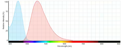
Fig. Fluorescence excitation and emission spectra of Hoechst 33342 bound to DNA
References:
[1]. Portugal J, Waring MJ. Assignment of DNA binding sites for 4′, 6-diamidine-2-phenylindole and bisbenzimide (Hoechst 33258). A comparative footprinting study. Biochimica et Biophysica Acta (BBA)-Gene Structure and Expression. 1988 Feb 28;949(2):158-68.
[2]. Weisblum B, Haenssler E. Fluorometric properties of the bibenzimidazole derivative Hoechst 33258, a fluorescent probe specific for AT concentration in chromosomal DNA. Chromosoma. 1974 Sep;46(3):255-60.
核酸染色剂Hoechst 33342 (Ex/Em: 350/461 nm) 常被用作细胞可渗透的核染色剂,与双链DNA结合后发出蓝色荧光。Hoechst 33342常被用于与细胞计数、细胞周期分析和细胞复制相关的各种研究中。它特别有用于鉴定凋亡细胞中的紧缩细胞核,并与BrdU染色结合在一起进行细胞周期研究。
Hoechst染料还可通过跟踪其发射光谱的变化来监测细胞活力。作为小沟结合DNA染料,具有AT选择性,Hoechst染料能够结合所有核酸,但与GC富集的双链DNA链相比,它们对富含AT碱基的链显示出更大的荧光增强[1]。这种特性已被利用于鉴定染色体中的Q带,这些区域富含AT碱基对,当用quinacrine染料染色时会发出明亮的荧光[2]。
实验参考方法 Experimental Reference Method
本方案仅提供一个指导,请根据您的具体需要进行修改。
1、制备Hoechst 33342染色液
(1) 配制Hoechst 33342染料储存液: 使用DMSO溶解固体Hoechst 33342,配置成10mg/mL的Hoechst 33342染料储存液。
注意: Hoechst储存液建议分装后于-4℃或-20℃避光保存,避免反复冻融。
(2)工作液制备:使用预热的无血清培养基或缓冲液(如HBSS或PBS)稀释储存液,配制浓度为10μg/mL的Hoechst 33342工作液。
注意: 请根据实际情况调整 Hoechst 工作液浓度,现用现配。
2、细胞染色
2.1 悬浮细胞(以6孔板为例)
(1)悬浮细胞经1000g离心3-5min。弃去上清液,使用PBS清洗两次,每次5分钟。
(2)加入1mL的Hoechst 33342染料工作液,室温避光孵育5-10 min分钟。
(3)孵育结束后,经1000g离心5分钟,去除上清液,加入PBS清洗2-3次,每次5分钟。
(4)使用无血清细胞培养基或PBS重悬细胞,通过荧光显微镜或流式细胞技术进行观察。
2.2 贴壁细胞
(1)在无菌盖玻片上培养贴壁细胞。
(2)从培养基中移走盖玻片,吸出过量的培养基,将盖玻片放在潮湿的环境中。
(3)从盖玻片的一角加入100uL的Hoechst 33342染料工作液,轻轻晃动使染料均匀覆盖所有细胞,室温避光孵育5-15min分钟。
(4)吸弃染料工作液,使用培养液洗盖玻片2~3次,通过荧光显微镜进行观察。
3.显微镜检测:Hoechst 33342的激发/发射光分别为350/460nm。
注意事项:
①对于固定的细胞或组织样品的染色,固定后需漂洗去除固定剂;
②Hoechst 33342染色通常在其他染色后进行,如果不需要进行其它染色,则直接进行Hoechst 33342染色;
③为减缓荧光淬灭,建议使用抗荧光淬灭封片剂;
④荧光染料均存在淬灭问题,请尽量注意避光;
⑤Hoechst 33342对人体有一定刺激性,为了您的安全和健康,请穿实验服并戴一次性手套操作。
References:
[1]. Lisa C Crowley, Brooke J Marfell , Nigel J Waterhouse. Analyzing Cell Death by Nuclear Staining with Hoechst 33342. 2016 Sep 1;2016(9). doi: 10.1101/pdb.prot087205.
产品文档 Product Documents
Purity:>98.00%
相关生物学数据Related Biological Data
1 / 2
化学性质Chemical Properties
CAS 号
23491-52-3
同义词
赫斯特荧光染料33342,bisBenzimide H 33342; HOE 33342
分子式
C27H28N6O
分子量
452.55 g/mol
溶解性
DMSO : 10 mg/mL (22.10 mM; Need ultrasonic)|Water : 1 mg/mL (2.21 mM; ultrasonic and warming and heat to 80°C)
保存条件
4°C, protect from light
General tips
请根据产品在不同溶剂中的溶解度选择合适的溶剂配制储备液;一旦配成溶液,请分装保存,避免反复冻融造成的产品失效。
储备液的保存方式和期限:-80°C 储存时,请在 6 个月内使用,-20°C 储存时,请在 1 个月内使用。
为了提高溶解度,请将管子加热至 37°C,然后在超声波浴中震荡一段时间。
储备液的保存方式和期限:-80°C 储存时,请在 6 个月内使用,-20°C 储存时,请在 1 个月内使用。
为了提高溶解度,请将管子加热至 37°C,然后在超声波浴中震荡一段时间。
Shipping Condition
评估样品解决方案:配备蓝冰进行发货。所有其他可用尺寸:配备 RT,或根据请求配备蓝冰。
计算工具摩尔浓度 / 稀释 / 分子量 / 单位换算 / 体内配方 / 溶解度
g/mol