


Orexin A (human, rat, mouse), orchestrates diverse central and peripheral processes[1]. Orexin A (human, rat, mouse) is a specific, high-affinity agonist for G-protein-coupled receptor OX1R. Orexin A (human, rat, mouse) has a role in the regulation of feeding behavior. Orexin A (human, rat, mouse) is an effective anti-nociceptive and anti-hyperalgesic agent in mice and rats[2].Orexins first bind OXRs, which in turn activate at least three subtypes of G-proteins (Gq/11, Gi/o, and Gs) or other proteins (e.g., β-arrestin). These effectors subsequently regulate phospholipases, ion channels, and protein kinases, ultimately triggering the activation of various downstream signaling pathways [3,5].
Orexin-A induced a transient increase in [Ca2+ ]i in CHO/OX1R cells in a dose-dependent manner, but failed to induce detectable [Ca2+ ]i transients in mock transfected CHO cells. The calcium mobilization is likely caused by the activation of the Gq class of heterotrimeric G proteins[6].The calculated concentration of orexin-A required to induce half-maximum response (EC50 ) was 30 nM. The concentration of unlabeled Orexin-A required to displace 50% of specific radioligand binding (IC50 ) was 20 nM. Repeated competitive radioligand binding assays and [Ca2+ ]i transient dose-response studies using stably transfected CHO cells expressing the human OX2R cDNA. The results demonstrated that OX2R is indeed a high-affinity receptor for human orexin-B, with an IC50 of 36 nM in the binding assay and an EC50 of 60 nM in the [Ca2+ ]i transient assay . Orexin-A had high affinity for this receptor, with 38 nM IC50 and 34 nM EC50 values.These findings confirm that orexin-A is indeed a specific, high-affinity agonist for OX1R.
The Orexin A concentration in cerebrospinal fluid (CSF) was abnormally low in seven of nine people with narcolepsy, implying that orexin transmission was deficient in these patients[7].In a later study, the same group reported a dramatic decrease in the CSF Orexin A levels in 32 of 38 successive narcolepsy-cataplexy cases [8].On the basis of these findings, they concluded that Orexin is deficient in most cases of human narcolepsy, suggesting possible diagnostic applications. Furthermore, the number of orexin neurons is reduced by 85%-95% in the LH of patients with narcolepsy [9]. Orexin mRNA and neuropeptide are completely absent in hypothalamus, pons and cortex of narcolepsy patients, and the secretory signal sequence of the orexin gene is deficient in the most serious cases of early onset narcolepsy [10].These observations further prove that narcolepsy is associated with deficiency in the orexin system.
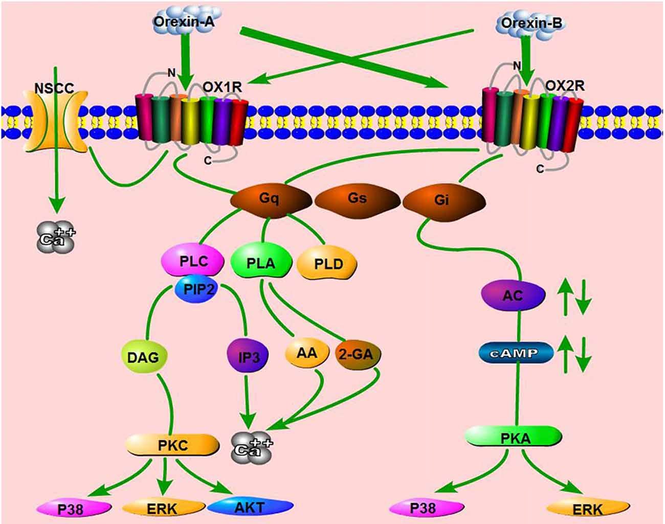
The binding of orexins to orexin receptor type 1 (OX1R) or OX2R stimulates Gq or Gi subtypes, which subsequently induce the activation of phospholipase C (PLC), phospholipase A (PLA), phospholipase D (PLD) or Adenylyl cyclases (AC), ultimately resulting in an increase in cytosolic Ca2+ and a downstream cascade response. In addition, OA binds OX1R and elevates Ca2+ by activating nonselective cation channels (NSCCs) [11].
References:
[1]. Bingham S, et al. Orexin-A, an hypothalamic peptide with analgesic properties. Pain. 2001 May;92(1-2):81-90.
[2]. Sakurai T, et al. Orexins and orexin receptors: a family of hypothalamic neuropeptides and G protein-coupled receptors that regulate feeding behavior. Cell. 1998 Feb 20;92(4):573-85.
[3].Dalrymple M B, Jaeger W C, Eidne K A, et al. Temporal profiling of orexin receptor-arrestin-ubiquitin complexes reveals differences between receptor subtypes[J]. Journal of Biological Chemistry, 2011, 286(19): 16726-16733.
[4].Kukkonen J P, Leonard C S. Orexin/hypocretin receptor signalling cascades[J]. British journal of pharmacology, 2014, 171(2): 314-331.
[5].Leonard C S, Kukkonen J P. Orexin/hypocretin receptor signalling: a functional perspective[J]. British journal of pharmacology, 2014, 171(2): 294-313.
[6].Hepler J R, Kozasa T, Gilman A G. [16] Purification of recombinant Gqα, G11α, and G16α from Sf9 cells[M]//Methods in enzymology. Academic Press, 1994, 237: 191-212.
[7].Nishino S, Ripley B, Overeem S, et al. Hypocretin (orexin) deficiency in human narcolepsy[J]. The Lancet, 2000, 355(9197): 39-40.
[8].Nishino S, Ripley B, Overeem S, et al. Low cerebrospinal fluid hypocretin (Orexin) and altered energy homeostasis in human narcolepsy[J]. Annals of Neurology: Official Journal of the American Neurological Association and the Child Neurology Society, 2001, 50(3): 381-388.
[9].Thannickal T C, Moore R Y, Nienhuis R, et al. Reduced number of hypocretin neurons in human narcolepsy[J]. Neuron, 2000, 27(3): 469-474.
[10].Peyron C, Faraco J, Rogers W, et al. A mutation in a case of early onset narcolepsy and a generalized absence of hypocretin peptides in human narcoleptic brains[J]. Nature medicine, 2000, 6(9): 991-997.
[11].Wang C, Wang Q, Ji B, et al. The orexin/receptor system: molecular mechanism and therapeutic potential for neurological diseases[J]. Frontiers in molecular neuroscience, 2018, 11: 220.
Orexin A(人类、大鼠、小鼠)协调不同的中枢和外周过程[1]。 Orexin A(人、大鼠、小鼠)是 G 蛋白偶联受体 OX1R 的特异性高亲和力激动剂。 Orexin A(人类、大鼠、小鼠)在调节摄食行为中发挥作用。食欲素 A(人、大鼠、小鼠)是小鼠和大鼠的有效抗伤害感受剂和抗痛觉过敏剂[2]。食欲素首先结合 OXRs,进而激活 G 的至少三种亚型-蛋白质(Gq/11、Gi/o 和 Gs)或其他蛋白质(例如,β-arrestin)。这些效应子随后调节磷脂酶、离子通道和蛋白激酶,最终触发各种下游信号通路的激活[3,5]。
Orexin-A 以剂量依赖性方式诱导 CHO/OX1R 细胞中 [Ca2+ ]i 的瞬时增加,但未能诱导可检测的 [Ca2+ ]i 模拟转染的 CHO 细胞中的瞬变。钙动员可能是由异源三聚体 G 蛋白的 Gq 类激活引起的[6]。计算的 orexin-A 诱导半数最大反应所需的浓度 (EC50) 为 30 nM。取代 50% 特定放射性配体结合所需的未标记 Orexin-A 浓度 (IC50 ) 为 20 nM。使用稳定转染的表达人 OX2R cDNA 的 CHO 细胞进行重复竞争性放射性配体结合测定和 [Ca2+ ]i 瞬时剂量反应研究。结果表明,OX2R 确实是人类 orexin-B 的高亲和力受体,结合试验中的 IC50 为 36 nM,EC50 为 60 nM在 [Ca2+ ]i 瞬时测定中。 Orexin-A 对该受体具有高亲和力,具有 38 nM IC50 和 34 nM EC50 值。这些发现证实 orexin-A 确实是一种特异性的、高- OX1R 的亲和激动剂。
9 名发作性睡病患者中有 7 人的脑脊液 (CSF) 中的食欲素 A 浓度异常低,这意味着这些患者的食欲素传递不足[7]。在后来的研究中,同一组报告称,连续 38 例嗜睡症-猝倒病例中有 32 例的 CSF Orexin A 水平显着下降[8]。根据这些发现,他们得出结论,Orexin A 水平在大多数人类病例中是缺乏的发作性睡病,提示可能的诊断应用。此外,发作性睡病患者 LH 中的食欲素神经元数量减少了 85%-95%[9]。发作性睡病患者的下丘脑、脑桥和皮质完全缺失食欲素 mRNA 和神经肽,并且在最严重的早发发作性睡病病例中食欲素基因的分泌信号序列缺失[10]。这些观察结果进一步证明发作性睡病与食欲素系统缺陷有关。
食欲素与食欲素受体 1 型 (OX1R) 或 OX2R 的结合刺激 Gq 或 Gi 亚型,随后诱导磷脂酶 C (PLC)、磷脂酶 A (PLA)、磷脂酶 D (PLD) 或腺苷酸环化酶 ( AC),最终导致细胞溶质 Ca2+ 增加和下游级联反应。此外,OA 结合 OX1R 并通过激活非选择性阳离子通道 (NSCC) [11] 来升高 Ca2+。