


MitoPQ (MitoParaquat) is a mitochondria-targeted redox cycler that selectively increases superoxide production within the mitochondrial matrix in vivo and in cells[1]. MitoPQ accumulates in the mitochondrial matrix and generates O2- through redox cycling at the flavin site of complex I (Fig. 1)[1,2]. MitoPQ is commonly used to study the role of mitochondrial superoxide production in health and disease in both cells and in vivo[3,4].
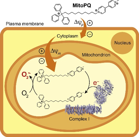
Fig. 1. Rationale for the development of MitoParaquat[1,2]. MitoParaquat (MitoPQ) is composed of a redox cycling paraquat moiety, and a hydrophobic carbon chain linking it to a mitochondria-targeting triphenylphosphonium cation. MitoPQ is accumulated by mitochondria driven by the plasma (Δψp) and mitochondrial (Δψm) membrane potentials. Within the matrix, the dicationic viologen component of MitoPQ is reduced to a radical monocation by one-electron reduction at the flavin site of complex I. The radical monocation then reacts very rapidly with O2 to generate superoxide. This localized redox cycling leads to the selective production of superoxide within the mitochondrial matrix.
In vitro, treatment of C2C12 myoblasts with MitoPQ (5μM) for 20min time-dependently and significantly increased MitoSOX fluorescence intensity, whereas paraquat (PQ) at equivalent conditions failed to increase MitoSOX oxidation within this timeframe. Treatment of HCT116 cells with MitoPQ (1-10μM) for 24h induced cell death in a dose-dependent manner, with significantly higher toxicity compared to PQ[1]. Treatment of Raw264.7 cells with MitoPQ (0.5μM) for 16h significantly disrupted the mitochondrial membrane potential, an effect that was attenuated by the addition of 10mM N-acetylcysteine (NAC)[5]. Treatment of 3T3-L1 adipocytes with MitoPQ (10μM) for 2h specifically increased mitochondrial superoxide and hydrogen peroxide levels without affecting global cellular respiration[6].
In vivo, acute intraperitoneal injection of MitoPQ (0.16mg/kg) into wild-type mice fasted for 16h significantly impaired glucose tolerance 2h post-injection. Intraperitoneal injection of MitoPQ (0.16mg/kg) in wild-type mice for 1.5h impaired hepatic insulin signaling in vivo, as evidenced by reduced levels of phosphorylated insulin receptor (IR), AKT, and GSK3α[7]. Daily intraperitoneal injection of MitoPQ (0.1mg/kg/day) for 7 days in cardiomyocyte-specific Nrf3 knockout mice significantly reduced survival rates and attenuated the beneficial effects of Nrf3 gene deletion on cardiac function and remodeling after myocardial infarction[8].
References:
[1] ROBB E L, GAWEL J M, AKSENTIJEVIĆ D, et al. Selective superoxide generation within mitochondria by the targeted redox cycler MitoParaquat[J]. Free Radical Biology and Medicine, 2015, 89: 883-894.
[2] COCHENÉ H M, MURPHY M P. Complex I is the major site of mitochondrial superoxide production by paraquat[J]. Journal of Biological Chemistry, 2008, 283(4): 1786-1798.
[3] ANTONUCCI S, MULVEY J F, BURGER N, et al. Selective mitochondrial superoxide generation in vivo is cardioprotective through hormesis[J]. Free Radical Biology and Medicine, 2019, 134: 678-687.
[4] GOLEVA T N, LYAMZAEV K G, ROGOV A G, et al. Mitochondria-targeted 1, 4-naphthoquinone (SkQN) is a powerful prooxidant and cytotoxic agent[J]. Biochimica et Biophysica Acta (BBA)-Bioenergetics, 2020, 1861(8): 148210.
[5] CHOWDHURY A R, ZIELONKA J, KALYANARAMAN B, et al. Mitochondria-targeted paraquat and metformin mediate ROS production to induce multiple pathways of retrograde signaling: A dose-dependent phenomenon[J]. Redox Biology, 2020, 36: 101606.
[6] FAZAKERLEY D J, MINARD A Y, KRYCER J R, et al. Mitochondrial oxidative stress causes insulin resistance without disrupting oxidative phosphorylation[J]. Journal of Biological Chemistry, 2018, 293(19): 7315-7328.
[7] GONCALVES R L, WANG Z B, RIVEROS J K, et al. CoQ imbalance drives reverse electron transport to disrupt liver metabolism[J]. Nature, 2025.
[8] CHEN Q, ZHENG A, XU X, et al. Nrf3-Mediated mitochondrial superoxide promotes cardiomyocyte apoptosis and impairs cardiac functions by suppressing Pitx2[J]. Circulation, 2025, 151(14): 1024-1046.
MitoPQ(MitoParaquat)是一种可选择性增加体内和细胞线粒体基质内超氧化物产生的线粒体靶向氧化还原循环剂[1]。MitoPQ 在线粒体基质中富集并通过复合物I的黄素位点的氧化还原循环产生O2-(图1)[1,2]。MitoPQ通常用于细胞或体内线粒体超氧化物产生在健康和疾病中作用的研究[3,4]。
图1. MitoPQ开发的基本原理[1,2]。MitoPQ由一个氧化还原循环的百草枯部分和将其连接到靶向线粒体的三苯基膦阳离子的疏水碳链组成。MitoPQ 由线粒体在血浆 (Δψp) 和线粒体 (Δψm) 膜电位的驱动下积累。在基质中,MitoPQ 中的双阳离子紫罗兰成分在复合物 I 的黄素位点通过单电子还原被还原为自由基单阳离子。然后,该自由基单阳离子与 O2 快速反应生成超氧化物。这种局部氧化还原循环导致线粒体基质内选择性产生超氧化物。
在体外,MitoPQ(5μM)处理C2C12成肌细胞20min,能时间依赖性地显著增加MitoSOX荧光强度,而同等条件下的Paraquat(PQ)在此时间范围内无法增加MitoSOX氧化。MitoPQ(1-10μM)处理HCT116细胞24h,能剂量依赖性地诱导细胞死亡,毒性远高于PQ[1]。MitoPQ(0.5μM)处理Raw264.7细胞16h,能显著破坏跨膜电位,而添加10mM N-acetylcysteine(NAC)可减弱MitoPQ的破坏作用[5]。MitoPQ(10μM)处理3T3-L1脂肪细胞2h,能特异性地增加线粒体超氧化物和过氧化氢,但不影响整体细胞呼吸[6]。
在体内,MitoPQ(0.16mg/kg)通过腹腔注射急性处理禁食16h的野生型小鼠,2h后显著损害了小鼠的葡萄糖耐受性。MitoPQ(0.16mg/kg)通过腹腔注射处理野生型小鼠1.5h,削弱了体内肝脏胰岛素信号传导(磷酸化胰岛素受体(IR)、AKT和GSK3α水平降低)[7]。MitoPQ(0.1mg/kg/day)通过腹腔注射处理心肌细胞特异性Nrf3敲除小鼠7天,小鼠的存活率显著降低,削弱了Nrf3基因缺失对心肌梗死后心脏功能和重塑的有益影响[8]。